
New article published on Scientific Reports by the Veterinarians of the Department of Animal Medicine, Productions, and Health of the University of Padua
Scientific reports is one of the most important open-access scientific journal currently available. Automatic classification of canine thoracic radiographs using deep learning is the new article released by the Veterinarians of the Diagnostic Imaging Unit of the University teaching Hospital of the Department of Animal Medicine, Productions, and Health (MAPS) of the University of Padua.
This paper describes a new algorithm, based on convolutional neural networks, capable of identifying some of the most common radiographic findings on canine thoracic radiographs. This algorithm is, prospectively, a useful tool to reduce the number of errors in the interpretation of thoracic radiographs by vets.
A Convolutional Neural Network (ConvNet/CNN) is a Deep Learning algorithm which can take in an input image, assign importance (learnable weights and biases) to various aspects/objects in the image and be able to differentiate one from the other.
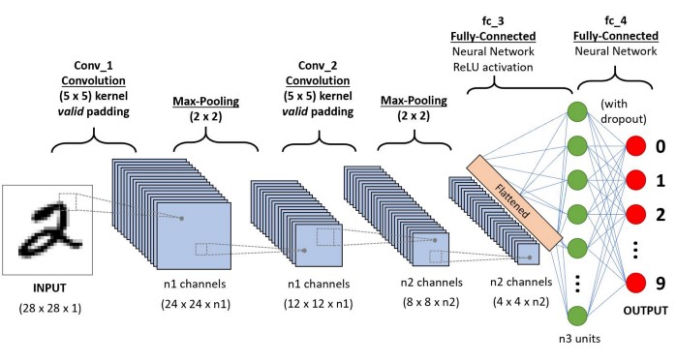
The architecture of a ConvNet is analogous to that of the connectivity pattern of Neurons in the Human Brain and was inspired by the organization of the Visual Cortex. Individual neurons respond to stimuli only in a restricted region of the visual field known as the Receptive Field. A collection of such fields overlap to cover the entire visual area.
The two main goals of this publication are:
- Develop an algorithm capable of detecting some of the most common lesions found on canine thoracic radiographs;
- Test the generalization ability of the developed algorithm on a different dataset.
To develop this algorithm the researchers of the MAPS department have collected and evaluated 3839 radiographs of the canine thorax (all acquired in a latero-lateral projection). The database was divided into two different dataset, on for the training and one for the testing of the algorithm.
The researchers have also developed a beta version of a website, called V.E.R.A. (Virtual Veterinary Radiology Assistant), where the vets can test the newly developed algorithm. In this website the vets can upload the radiographs (in a DICOM format) and directly visualize the results of the analysis.
For more information regarding V.E.R.A. (Virtual Veterinary Radiology Assistant) please contact dr. Tommaso Banzato (Tommaso.banzato@unipd.it)